Issues with the heart can take many forms. Your cardiologist may order a combination of tests to help diagnose your issue. Below are some common tests:
Electrocardiogram (ECG or EKG) – an electrocardiogram records the electrical signals of your heart. In the doctor’s office or hospital, sensors are attached to the chest that record the hearts electrical signal.
Holter Monitor – a small wearable device that tracks all your heartbeats and records your heart rhythm. It may be worn for one or two days.
Echocardiogram (Echo) – an echocardiogram uses sound waves to produce an image of your heart. Cardiologist can see your heart beating and pumping blood. There are several different types of these tests:
Transthoracic echocardiogram – a technician guides a transducer that is pressed firmly against your chest across it. This records sound waves which a computer converts to an image.
Transesophageal echocardiogram – sometimes it is you doctor needs a more detailed image or the image is hard to capture. In this case, a transducer is inserted in the esophagus after the throat has been numbed.
Learn more about Transesophageal Echocardiogram
Doppler echocardiogram – doppler technique can be added to an echo to measure the speed and direction of blood flow.
Stress echocardiogram – Some heart issues only show up during exercise. During a stress echo, ultrasound images are taken of the heart before and during a walk on a treadmill. If you are unable to exercise a medication called dobutamine can be used to make the heart beat faster.
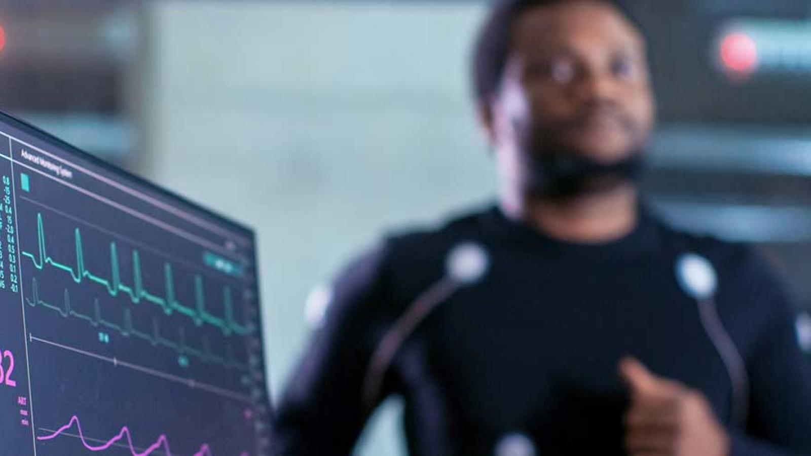
Cardiac Stress Test (Exercise Stress Test) -- the heart’s activity is monitored during physical activity. While you walk on a treadmill or ride a stationary bike, your heart rhythm, blood pressure and breathing are monitored.
Nuclear Stress Test – radioactive dye in the heart is used to create an image of the heart before and during exercise.
Cardiac Catheterization – a small thin tube called a catheter is threaded through a vessel in the groin, neck or wrist to the heart.
Angiogram uses x-ray imaging to see the blood vessels in your heart. Dye is injected into the heart or blood vessel through a catheter. The dye is visible on x-ray. Cardiologists look for restricted vessels and can also open clogged vessels during an angiogram.
Chest X-ray – a picture of the heart, lungs, and bones of the chest. It is used to determine if the heart is enlarged or if the lungs have fluid in them.
Call 866-GUTHRIE to schedule a testing appointment.
Cardiac Testing at Guthrie
- We offer diagnostic testing in 15 locations in New York and Pennsylvania. Our team uses the latest techniques and advanced diagnostic imaging equipment to accurately diagnose cardiac and vascular disease.
- Board-certified cardiology specialist: Guthrie non-invasive and interventional cardiologists, electrophysiologist and cardiac, thoracic and vascular surgeons are specially trained in cardiovascular care and board certified.
- Guthrie Heart and Vascular Care Center, located in Guthrie Robert Packer Hospital, is a state-of-the-art space for heart and vascular testing and treatment. Located near the emergency department and ICU, there are private patient rooms, cardiac catheterization labs, and a hybrid operating room. The unit is centered around quick efficient care of our patients.


